2025년 5월 21일 수요일

Zygoma Reconstruction Surgery


 668. <Zygoma Reconstruction Surgery>

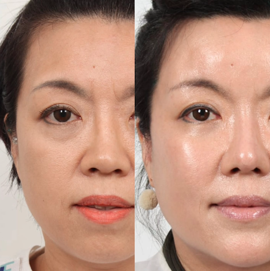
Zygoma reconstruction is performed when the cheekbone (zygoma) is deformed due to trauma, tumor removal, congenital conditions, or previous surgeries. Severe cases may require repositioning the bone or placing an artificial implant to restore symmetry and structure.

Since the zygoma supports the orbit, its collapse can lead to enophthalmos (sunken eye). Therefore, correcting both the bone and the orbit is often necessary. Advanced techniques like 3D-printed implants, mirroring of the unaffected side, and navigation systems help ensure precision. Surgical guides may also be used for accurate implant placement.

When synthetic implants aren't preferred, autologous tissues like jawbone (from mandibular contouring), rib, or skull can be used. In milder cases, microparticulated costal cartilage mixed with fibrin glue can reconstruct the area like sculpting clay.

Zygoma reconstruction is more than cosmetic—it restores facial structure and function. Preoperative checks, especially for sinus health, are essential to avoid complications.

[It’s a reconstructive surgery to restore midface depression.]

— 668mm Growing Pine Tree —

2025년 5월 19일 월요일

Zygoma Augmentation Surgery


667. <Zygoma Augmentation Surgery>

Zygoma augmentation surgery is a cosmetic surgery performed to increase the volume of the zygomatic (cheekbone) area when the midface region is underdeveloped. It is mainly found among Westerners, where the zygomatic bone area is less developed, making the midface appear flat and lacking volume, prompting the surgery to add dimensionality.

The zygomatic bone is a structure that extends from the center of the face to the side, playing an important role in determining the contour and three-dimensionality of the face. In East Asians, it is relatively more developed, while in Westerners, it is relatively less developed. There are various reasons for this, but one explanation is that East Asians tend to have a brachycephalic (short-headed) skull shape, while Westerners tend to have a dolichocephalic (long-headed) skull shape. Evolutionarily, due to a more collectivist development, East Asians tend to follow commands more and prioritize the species' standpoint, leading to temporal lobe development and suppression, activating a psychological mechanism to seek pleasure through habits like teeth clenching. This results in more developed zygomatic bones and lateral skull areas. On the other hand, Westerners, due to individualistic development, tend to think more, talk more, and behave more freely, which leads to frontal lobe development and thus more developed forehead regions with less developed zygomas.

To undergo zygoma augmentation surgery, preoperative planning is necessary. Through a 3D CT scan, the facial bone structure, asymmetry, presence of maxillary sinus abnormalities, and zygoma protrusion direction are carefully examined. Using a 3D printer, a customized implant is created to enlarge the zygomatic bone. At this time, care must be taken to ensure that the infraorbital nerve is not compressed when inserting the implant, by leaving a gap to prevent nerve pressure. The implant is inserted above the bone through an incision inside the mouth, thereby making the midface more three-dimensional.

Zygoma augmentation may also be considered due to age-related midface volume loss, or when the zygomatic bone is depressed due to trauma. In such cases, the zygoma can be osteotomized and elevated, and both the frontal and temporal limbs of the zygoma are osteotomized and fixed with titanium.

If the depressed bone is too difficult to elevate through osteotomy, artificial implants can be inserted over the maxilla and zygoma. Especially in trauma-related cases, if the maxillary sinus is fractured and sinusitis is present, inflammation could spread to the implant, so preoperative management and control of rhinitis or sinusitis are required. If one wants to avoid complications like infection from artificial implants, autologous tissue grafts can be used as an alternative. However, since there are size limitations with autologous implants, they may not be suitable for significantly augmenting the zygoma. One method involves grafting the mandibular bone (obtained while slimming the jaw) onto the zygoma. For a more ergonomic bone graft, bone particle grafting can be performed by grinding the bone. However, in the case of bone particle grafts, the absorption rate can exceed 30%, and inflammation may occur in some cases, so the physical condition must be carefully checked before surgery.

Fat grafting is the best way to minimize inflammation risk while using autologous tissue, and can be used as an alternative to elevate the zygoma. If only a slight augmentation is needed, costal cartilage can be harvested and cartilage particle grafting can be partially injected to slightly enhance the zygoma.

Thus, zygoma augmentation can be performed using either artificial or autologous tissue to provide volume to the midface.

[Zygoma augmentation surgery is a cosmetic surgery that enhances the three-dimensional appearance of the midface.]

– 667mm Growing Pine Tree –

Zygoma Reduction Surgery


 666. <Zygoma Reduction Surgery>


Zygoma reduction surgery reduces prominent cheekbones for a softer contour and slimmer mid-face. The procedure involves intraoral incisions and L-shaped osteotomy to push and fix the zygoma inward, adjusting both position and size.

There are two types: incision and non-incision for the zygomatic arch. The non-incision method avoids sideburn scars but skips arch fixation, which can cause bone nonunion — especially in patients with teeth grinding or clenching habits. For them, dual fixation via incision is safer, though a small scar remains near the sideburn.

In patients over 40, reduced bone volume without skin reduction can lead to sagging. A facelift performed together can prevent this, using one incision for both surgeries and leaving no additional scars.

Facial asymmetry from chewing or smiling habits requires custom planning. Arch incisions must also avoid facial nerve damage that may affect eyebrow movement.

Post-op, jaw stiffness may occur. Mouth-opening exercises are important. Non-surgical methods like thread lifts or fat removal rarely reduce actual bone. For real contour change, bone surgery is necessary.

Zygoma reduction improves both side and 45° angles but may create a visible second curve. It can also make the jawline appear more prominent, so considering jaw surgery together may be ideal.

[Zygoma reduction narrows the mid-face width.]
— 666 mm Growth Pine Tree

Quick Zygoma Reduction Surgery


 665. <Quick Zygoma Reduction Surgery>

Quick zygoma surgery is promoted as a less invasive, faster-healing alternative to traditional cheekbone reduction. It replaces general anesthesia with sedation, reduces surgical time, and minimizes scarring — leading to the perception that it's an “easier” option.

One common method involves fracturing and pushing in only the zygomatic arch, without cutting into the main zygoma body. This can be done through incisions inside the mouth, in front of the sideburn, or hidden in the hairline. While this narrows the face from the side, it doesn’t reduce the 45-degree angle (the front-side cheekbone), which can lead to disappointment for those with both prominent side and 45° cheekbones. In such cases, the 45° cheekbone may appear even more pronounced after surgery.

Issues like nonunion, over- or under-correction, and bone relapse are not uncommon, especially when fixation is skipped. If revision is needed, the zygomatic arch must often be restored before performing full reduction again — turning a “simple” surgery into two major ones.

Quick methods may seem convenient but often have limited effects and apply to only a narrow group of patients. If done without precise planning and post-op care, they can become a source of dissatisfaction.

New doesn’t always mean better. If cheekbone reduction is truly needed, a full and properly planned surgery by an experienced surgeon — not just a faster one — is the best route.

[Quick zygoma surgery is just a quick partial cheekbone reduction.]

— 665 mm Growth Pine Tree

Buccal Fat Removal: What Are You Really Removing?


 663.Buccal Fat Removal: What Are You Really Removing?

Buccal fat removal is a cosmetic surgery designed to slim the face by removing the buccal fat pad — a deep fat compartment located between the cheek and the upper jaw muscles. This pad stretches in four directions, divided into three lobes, reaching from the parotid gland in the front to the temporalis muscle and mandibular ramus in the back.


But why does buccal fat exist?

Fat is our body’s form of energy storage. We store and use energy through three main pathways: sensory, cognitive, and motor.


Motor energy is stored in abdominal fat — our belly reflects our movement energy.


Sensory energy is stored in facial fat, especially buccal fat.


The brain (hippocampus, frontal lobe) processes cognitive energy.


Think of the buccal fat pad as a storage unit for sensory energy.

When you overuse your senses or are mentally stressed, your cheeks hollow out — you look tired or gaunt. But when you feel safe, calm, and less stimulated, your cheeks fill out naturally.


Trying to gain buccal fat by just eating more? You'll only gain belly fat instead. Want to lose cheek fat? Increase sensory input and anxiety — though that's not advisable.

The point is: Buccal fat changes naturally depending on how you live and feel.


So what happens when you surgically remove it?

You’re permanently discarding a reservoir of sensory energy. Over time, this may lead to faster eye fatigue, earlier onset of tinnitus, and even reduced alertness. As we age, we naturally use more sensory energy — removing the storage when you’re young could leave you drained later in life. Once it’s gone, it’s nearly impossible to restore.


Before choosing this surgery, consider:

Are you paying to destroy your own sensory reserve — your deep emotional storage — with trauma disguised as beauty?

This isn’t just a cosmetic decision. It’s an energetic one.


[Buccal Fat Removal = Cutting Away Your Sensory Energy Bank]

— 663mm Growth Pine Tree 🌲


#buccalfatremoval #facecontouring #innerhealth #aesthetictruth #deepbeauty

2025년 5월 14일 수요일

Buccal Fat Removal Surgery


663. <Buccal Fat Removal Surgery>

Buccal fat removal is a cosmetic procedure that removes or reduces the buccal fat pad—located deep between the cheek and the upper jaw—to create a slimmer facial contour. The buccal fat pad lies between the buccinator and zygomatic muscles and extends in four directions, with its front near the parotid gland and rear toward the temporalis muscle and mandible.

Why does this fat exist? Fat tissue stores energy, and our body uses energy in three ways: through movement, thinking, and sensory activities. Belly fat stores energy for movement, especially leg use. Cheek fat—specifically buccal fat—stores energy for sensory functions such as sight, hearing, smell, taste, and touch. Visual energy is stored in the orbital fat around the eyes, while general sensory energy is stored in the cheeks.

This means when you overuse your senses or feel stressed, your cheek fat may reduce, making your face look slimmer. If you're mentally relaxed and use fewer sensory functions, cheek fat tends to increase. While belly fat increases from overeating and inactivity, cheek fat increases from emotional comfort—not food. Trying to gain cheek fat by eating more often just increases belly fat. To add cheek volume, calm your mind. To reduce it, stress alone may work—no surgery needed.

So what happens if you surgically remove buccal fat? You're cutting out your sensory energy storage. Over time, this can lead to faster eye fatigue, early tinnitus, and reduced alertness. As we age, we naturally rely more on sensory input, depleting these reserves. Removing buccal fat while young erases what might be hard to restore later. Even though the surgery is relatively simple—done through an incision inside the mouth—it’s important to weigh the long-term effects before proceeding.

Consider whether it’s truly worth paying to destroy such a meaningful energy reservoir. Reflect deeply on the true purpose and impact of buccal fat removal.

[Buccal fat removal is the removal of your sensory energy reservoir.]
663mm Growing Pine Tree

#BuccalFatRemoval #SlimFaceSurgery #CheekFatReduction #YouthfulLook #FacialContouring #MidFaceLifting #PreventSagging #EnergizedFace #EpillPlasticSurgery

2025년 5월 13일 화요일

Cheek Fat Grafting


662. <Cheek Fat Grafting>

Cheek fat grafting is a cosmetic procedure that involves transplanting fat into the cheek area to improve sunken cheeks and enhance the volume and three-dimensional contour of the midface.

There are two types of cheek fat: superficial and deep. First, the superficial fat, known as the malar fat pad, is located above the orbicularis oculi muscle, sitting atop the zygomatic bone under the eyes. It plays a key role in shaping the midface. The malar fat pad is divided into compartments, including the infraorbital compartment, medial cheek compartment, and nasolabial compartment. These compartments are separated by the medial cheek septum and the nasolabial septum, respectively.

Second, the deep fat, called the buccal fat pad, lies between the buccinator and zygomaticus muscles. It consists of three lobes that extend in four directions. The anterior lobe surrounds the parotid gland, the intermediate lobe lies between the anterior and posterior lobes, and the posterior lobe extends toward the temporalis muscle and mandibular ramus.

The buccal fat pad serves as a reservoir for sensory energy. While the orbital fat inside the orbicularis oculi within the orbital cavity acts as an energy reservoir for vision, the buccal fat pad in the oral cavity stores energy for general sensory functions. When we eat a lot, we use the buccinator muscle, reducing the buccal fat above it; similarly, frequent chewing activates the temporalis muscle, depleting the deep fat overlying it. However, during periods of rest, when these muscles are not in use, energy is replenished and stored in the cheek fat.

On the other hand, if a person experiences high levels of stress, overuses visual and auditory senses, speaks excessively, loses appetite, and avoids eating due to aversion to smells, energy is continuously depleted without being replenished. This can lead to progressive fat loss in the cheeks, resulting in a gaunt, skeletal appearance over time.

Conversely, if someone eats well and feels relaxed, cheek fat tends to increase. Another deep fat layer is the suborbicularis oculi fat (SOOF), located beneath the orbicularis oculi muscle in the lower eyelid. It acts as a cushion and maintains the soft contour and volume around the eyes, contributing to a youthful and healthy look. When SOOF diminishes or sags, the under-eye area may appear hollow, highlighting dark circles and forming tear troughs—common signs of aging.

Thus, if a patient has prominent tear troughs, fat is grafted into the SOOF area. If the area in front of the cheekbone looks flat, and the zygomatic bone itself is under-projected, fat is evenly injected into the periosteum, muscle, and subcutaneous fat layers of the cheekbone region.

Fat for grafting is usually harvested from the abdomen or thighs. It is then refined using a centrifuge and loaded into a fat injector. For precision, a micro-fat injection device is used, allowing for injections as small as 1/240cc. Typically, amounts between 1/120cc and 1/60cc are injected to balance cheek volume symmetrically. For example, if one has a habit of smiling more on one side, the buccinator muscle on that side may be slightly thicker. Therefore, more fat is injected on the side with the lower mouth corner, and less on the lifted side, to achieve better symmetry. Rather than injecting equal amounts, the procedure accounts for bone recession, muscle thickness, and soft tissue depth for meticulous adjustment.

For secondary grafts, patients may opt for cryopreserved fat that can be thawed and reinjected 2–3 months later. However, the fat's freshness declines, leading to a slightly higher absorption rate. This factor should be considered when planning a second fat graft.

Most patients seeking cheek fat grafting tend not to smile often, so practicing smiling to engage and lift the buccinator muscle is recommended.

[Cheek fat grafting replenishes the energy reservoir of the senses and restores aesthetic volume.]

—662mm Growth Pine Tree—